Mediastinal lymphadenopathy in congestive heart failure
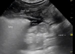
It’s an observation of ours that mediastinal lymph nodes are often prominent in cats with congestive failure. This can cloud the issue of identifying the primary cause of pleural effusions.
I haven’t come across a published account of this phenomenon although I doubt it’s a completely novel observation. In human medicine it’s an established feature:
Congestive Adenopathy: A Mediastinal Sequela of Volume Overload.
Shweihat YR, Perry J, Etman Y, Gabi A, Hattab Y, Al-Ourani M, Santhanam P, Bartter T.
J Bronchology Interv Pulmonol. 2016 Oct;23(4):298-302.
https://www.ncbi.nlm.nih.gov/pubmed/27623420
Mediastinal lymphadenopathy in congestive heart failure: a sequential CT evaluation with clinical and echocardiographic correlations.Chabbert V, Canevet G, Baixas C, Galinier M, Deken V, Duhamel A, Otal P, Joffre F, Remy J, Remy-Jardin M.Eur Radiol. 2004 May;14(5):881-9
https://www.ncbi.nlm.nih.gov/pubmed/14689226
An example: a feline patient of ours from last week:

The mediastinal nodes are large enough to raise interest in the possibility of, for example, lymphoma: were it not for the fact that there is good evidence for CHF. Not only is there pleural space disease (effusion). The lung itself is also markedly abnormal. This is best seen in video:
Normal ‘A lines’ parallel to the pleura are obscured by coalescing ‘B lines’ perpendicular to the lung margin….and echocardiographic findings are consistent with congestive failure.
I’ll spare you the details but, essentially, biatrial dilation, Doppler findings consistent with LV restrictive filling pattern and lack of hypertrophy are all consistent with restrictive cardiomyopathy.
We don’t have cytology or histopathology to confirm what’s going on in those nodes. But we’ve seen a few similar cases -enough to have a strong suspicion that enlarged lymph nodes shouldn’t necessarily dissuade us from a diagnosis of CHF.


