cardiology
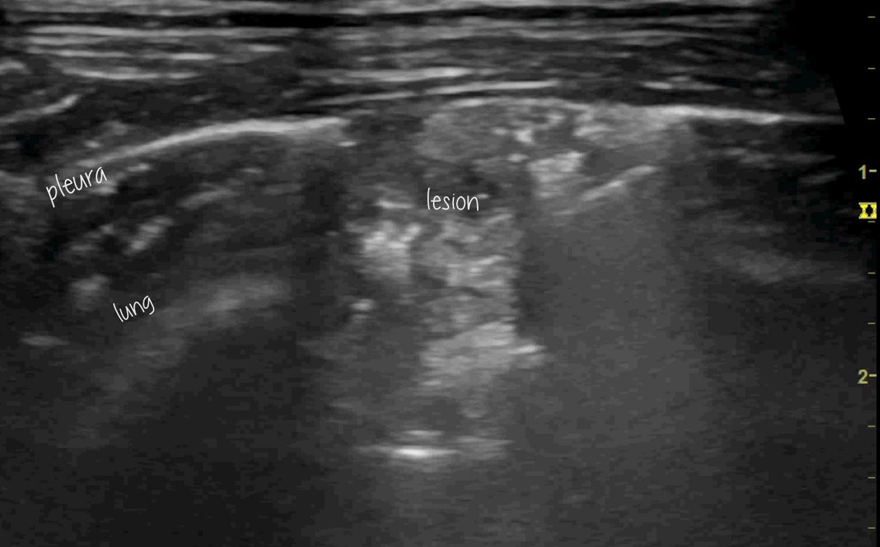
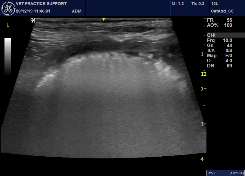
Lung ultrasound in the diagnosis of presumed pulmonary mycobacteriosis in a cat
Surprisingly, considering the wealth of information on conventional bacterial pneumonia, there is little published on the features of pulmonary TB in people. Int J Environ Res Public Health. 2018 Oct; 15(10): 2235. Potential Diagnostic Properties of Chest Ultrasound in Thoracic Tuberculosis—A Systematic Review Francesco Di Gennaro,1,2,† Luigi Pisani,3,4,† Nicola Veronese,5 Damiano Pizzol,6,* Valeria Lippolis,4 Annalisa … Continue reading
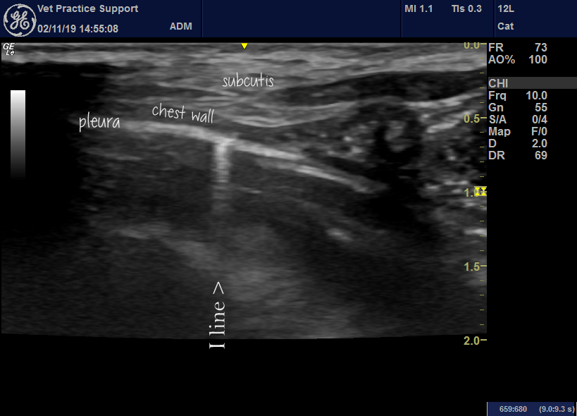
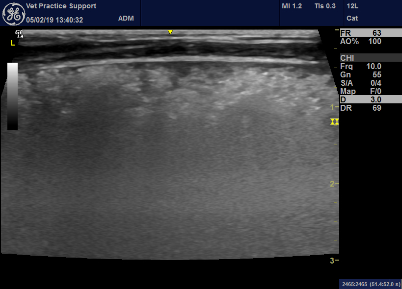
Lung ultrasound: contusions, interstitial syndrome, A, B, C, E, I and Z-lines explored
I reckon lung ultrasound must be one of the most profound developments in veterinary medicine during my career. The facility to diagnose rapidly, and with high degree of confidence, pneumonia, pulmonary oedema, contusions, pneumothorax and neoplasia is a phenomenal advance. Having arrived in our tool-kit in the last 10 years, this is still an evolving field. In this … Continue reading
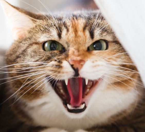
Gabapentin as a sedative/anxiolytic prior to ultrasonography in cats
Gabepentin is an enormously useful drug to facilitate ultrasonography in cats. I’m sure most of you are aware of this by now but it’s worth publicising a little more because, to get best effect, the use of gabapentin needs to be planned in advance of the day of scanning. J Am Vet Med Assoc. … Continue reading
Do we still believe that the EPIC study was helpful? When are we using pimobendan in dogs with mitral valve disease in early 2019?
Time has moved on a bit. We’ve had the benefit of a chance to reflect on EPIC: J Vet Intern Med. 2016 Nov;30(6):1765-1779. doi: 10.1111/jvim.14586. Epub 2016 Sep 28. Effect of Pimobendan in Dogs with Preclinical Myxomatous Mitral Valve Disease and Cardiomegaly: The EPIC Study-A Randomized Clinical Trial. Boswood A et al. https://onlinelibrary.wiley.com/doi/full/10.1111/jvim.14586 The … Continue reading
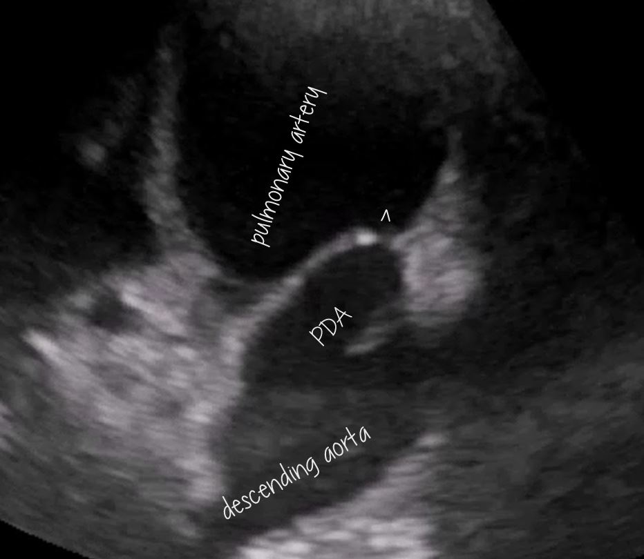
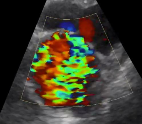
Ultrasonographic features of acute canine lung lobe torsion
It’s been a while since I’ve had a chance to scan a lung lobe torsion case: since when my machine got upgraded. This patient is a young Whippet with a 24 hour history of acute-onset cough, tachypnoea and pleural effusion (modified transudate). Her echocardiogram is interesting. Although we have to take into account the fact … Continue reading
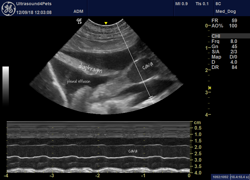
Iatrogenic overhydration: diagnostic nightmaresville
The inconvenient bottom line is that there is nothing intrinsic about the appearance of effusions or tissue oedema caused by fluid overload to distinguish them from those caused by congestive heart failure or any other pathological process. We have to infer the probable cause (or causes) from history, physical examination, clin path and imaging. So, cardiogenic … Continue reading
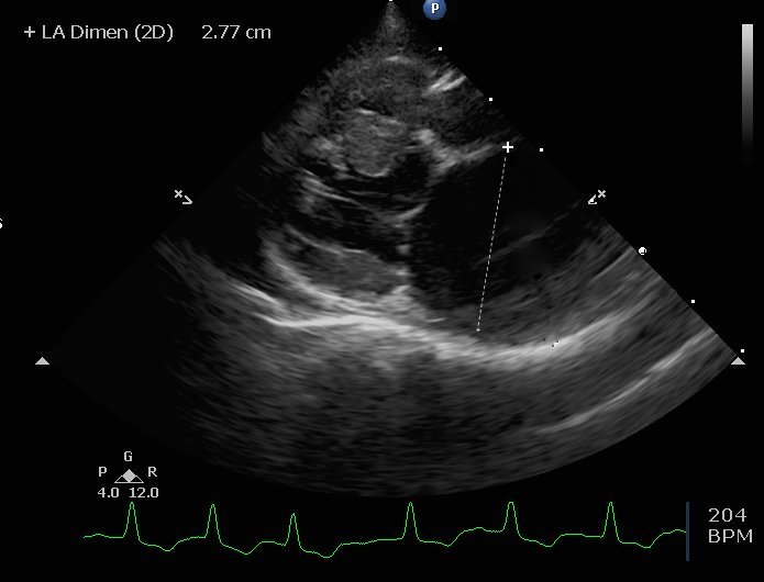
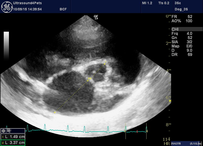
Why is this cat with a huge left atrium not in congestive failure?
This is a cat we saw last week. She presented for routine vaccination and was found to have a tachydysrhythmia. No hyperpnoea, no dyspnoea, outwardly well. This is her echocardiography. First, right long axis four chamber view: Her left atrium measures about 28mm (normal being <16mm). Technically, that falls into the ‘huge’ category. … Continue reading


