Anal sac ultrasound in dogs: a case of emphysematous anal sacculitis
Amazingly (actually maybe it’s not amazing :/) there appears to be just one published study on ultrasonography of canine anal sacs. This reflects a general lack of enthusiasm for anyone getting to grips with pathological conditions of the AGs. And it’s a sad state of affairs since they’re easy and rewarding to scan.
That one ground-breaking study is:
Diagnostic imaging features of normal anal sacs in dogs and cats
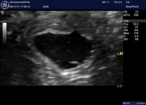
Technique is straightforward: a linear probe gives best results and i usually scan in a transverse plane from a point ventral to the anus with the beam directed dorsally and about 20 degrees angled cranially.
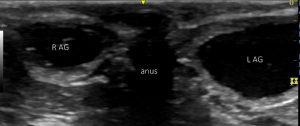
Transverse plane view of the anal glands from a point in the midline below the anus with the beam directed slightly cranially.
Simples.
The present patient has a history of chronic anal irritation unresponsive to various antibiotics, glucocorticoids or oclacitinib.
On sonography of the perianal area:
This is really not normal. The left anal gland is OK but on the left the anal gland lumen is gas filled.
In video:
Cytology of right anal gland secretion revealed large numbers of degenerate neutrophils and bacteria: some of which were intracellular.
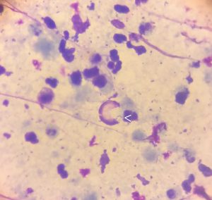
AG content cytology: intracellular cocci are arrowed
About 10% of ‘normal’ asymptomatic dogs have neutrophils in their anal gland secretions. However, they tend to be <10 per HPF and non-degenerate:
J Vet Med A Physiol Pathol Clin Med. 2004 Jun;51(5):249-53.
Gross and cytological characteristics of normal canine anal-sac secretions.
At the time we theorised that this might be an emphysematous anal sacculitis (not that such a thing has ever been reported to the best of our knowledge…but it seemed like a reasonable explanation). On this basis 3 weeks of co-amoxiclav were dispensed: to no noticeable effect.
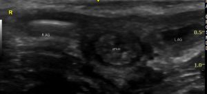
Although, on re-examination post-antibiotic the gas had vanished from the right gland:
R gland, transverse view, post-antibiotic
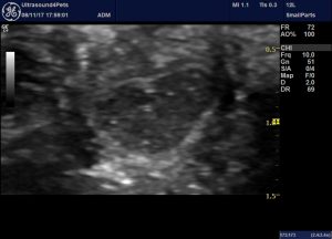
However, the perisaccular soft tissue remains patchily hyperechoic and the contents are hyperechoic compared to the left gland:
Left anal gland, transverse plane, post-antibiotic
Bilateral anal sacculectomy was performed. Histopathology:
‘The epithelial lining of the sac is extensively ulcerated and there is a mixed inflammatory reaction surrounding the wall with occasional lymphoid follicles’
On culture a mixed growth was present in samples from both glands. That from the right gland featured a heavy growth of Staph pseudintermedius sensitive to most antibiotics including co-amoclav.
This is all interesting on several fronts: firstly, I can’t find any other reports of ultrasonographic appearance of anal sacculitis in dogs. Secondly, emphysematous anal sacculitis may be a previously-undescribed entity. I find it easy to imagine that an anal gland full of gas might be painful. Thirdly, a 3 week course of an apparently appropriate antibiotic filed to eliminate an apparently sensitive Staph from the anal gland.


Your article is wonderful! I’m a surgeon trying to locate both anal sacs after another vet did a “sedate/flush” and follow up vets say both sacs are “gone” but duct remains and ends in a deep perianal open tract that appeared one month after flush. Because of you, I will try to ultrasound to determine if sacs are truly gone. Any thoughts?
7 yr MN eng bulldog
Dr. Julie Banks
Hi Julie, yes that’s a really good plan I think. It would be useful to know whether the inflammation looks focussed on the AGs. To be honest I certainly wouldn’t rule out anal furunculosis even though this isn’t a typical breed. I’d be inclined to assess sonography and get some tissue for histopathology because if AF then worth considering ciclosporine either as alternative to surgery or before sacculectomy. I find pathologists sometimes wary of committing to a diagnosis of AF but findings are usually strongly suggestive even if not definitive. https://doi.org/10.1111/j.1748-5827.1992. tb01062.x
Thank you very much for your article! Congrats from Brazil! it would be awesome if we could exchange ideas about US!
You’re very kind!thank you…. It’s just nice if anyone reads them!!
Dog 13 yrs old with enlarged anal sac. The degree of anisocytosis and anisokaryosis is mild to moderate. Acinar formation seen. Since mild to moderate, would this be cancer? What would be the life expectancy for this dog?
Sorry, I’m late replying. Obviously it’s difficult to comment on your specific case…i’m not a pathologist. But, any anal sac mass is a big concern as most are carcinomas. Life expectancy of any 13 y.o. dog is dependent on breed and any comorbidities and those are important considerations in deciding what to do about the AG lesion.