Tag Archives: sonography
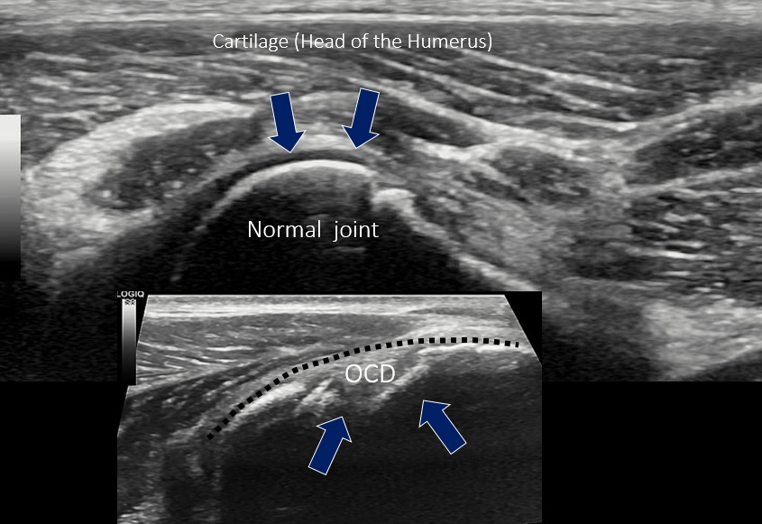
Sonographic features of septic arthritis in the dog
This is a nice article from a few years back: BMJ. 2014 May 29;348:g3463. doi: 10.1136/bmj.g3463. Will ultrasound scanners replace the stethoscope? Wittenberg M. https://www.bmj.com/content/348/bmj.g3463 The crux of it being that human medics are finding more and more advantages of using point-of-care ultrasound (POCUS) more as part of the physical examination, like a stethoscope, … Continue reading
Sonography of central line catheter infection in a dog
My understanding is that sonography is increasingly used in human medicines to guide interventions such as vascular access. This is a critical care case for which we were asked to perform thoracic and abdominal sonography. However, the most immediate problem turned out to be sepsis stemming from the jugular vein central line. &nbs… Continue reading
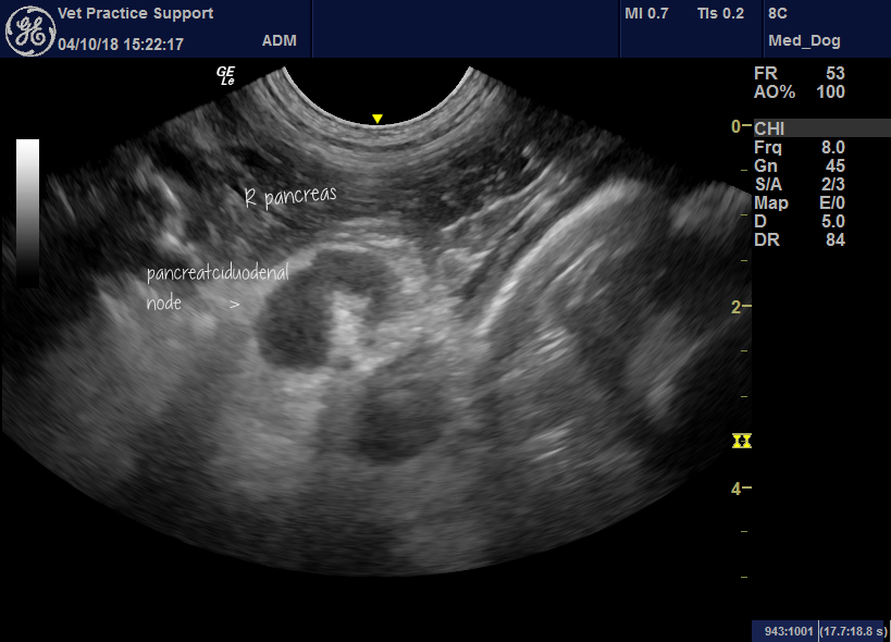
Sonographic appearance of pancreaticoduodenal lymph nodes in dogs and cats
Sonographic heterogeneity of abdominal lymph nodes has been reported as a feature associated with malignancy in dogs: Vet Radiol Ultrasound. 2007 Nov-Dec;48(6):565-9. Association between malignancy and sonographic heterogeneity in canine and feline abdominal lymph nodes. Kinns J1, Mai W. https://www.ncbi.nlm.nih.gov/pubmed/18018731 In that series, 91% of dogs with heterogeneous abdominal nodes proved to have malignancies. … Continue reading
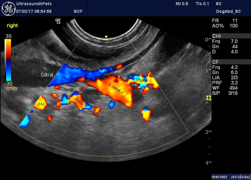
Multiple acquired portosystemic shunts (MAPSS) in dogs due to portal hypertension are not necessarily accompanied by hepatofugal flow in the normal tributaries of the portal vein
This is an issue I used to wonder about. When you see acquired shunts running, for example, from splenic to left renal veins….should flow in the gastrosplenic vein also be hepatofugal? That is, does blood flowing down the mesenteric vein from the guts continue into the main portal vein or does it all get diverted … Continue reading
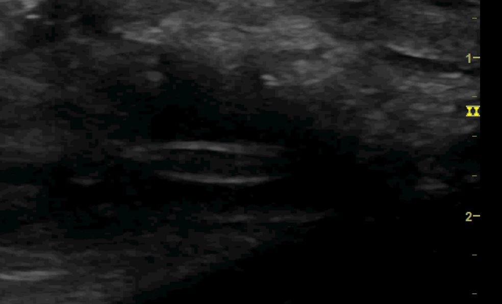
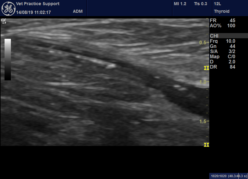
Grass seed foreign bodies: sonography and ultrasound-guided retrieval
Grass seed (and other vegetation) foreign bodies are a potential nightmare. We all know this. Sadly, it seems that neither CT nor MRI are reliable modalities for locating them in our patients. Obviously there may be clues from advanced imaging findings but actual visualisation of the foreign body itself isn’t consistent. In a series of … Continue reading
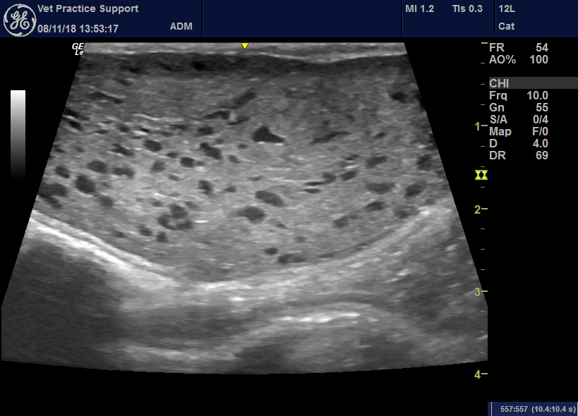
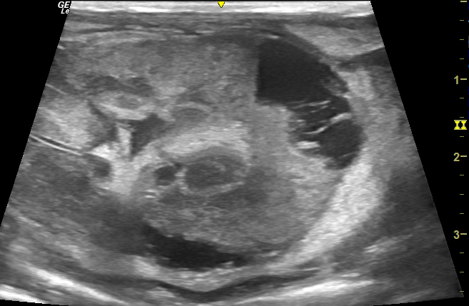
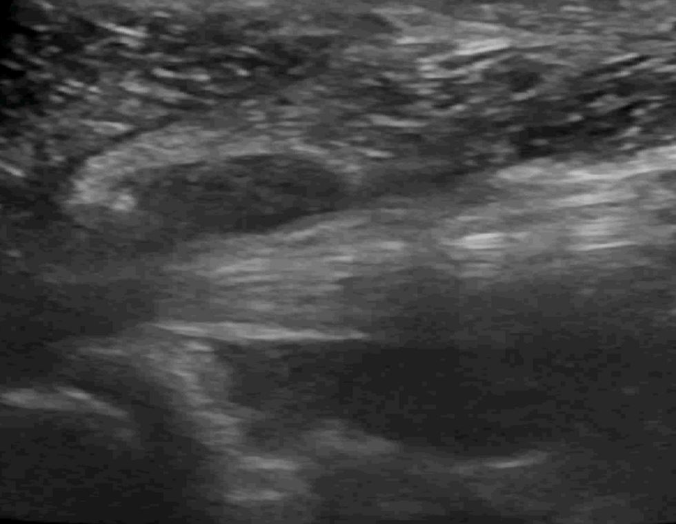
Sonography of canine mammary glands during lactation
Just a striking image of something not much depicted in the published literature. This is what normal, healthy lactating mammary gland looks like in the bitch (on this occasion two days post partum): &nbs… Continue reading


