

How should we manage dogs with mitral valve disease close to the B1/B2 boundary?…and related questions.
I should probably say at the outset that my views expressed here are my own. Within our team we have a healthy spectrum of opinion.
The concept of stage B2 heart disease is simple enough: we started with the idea that these are animals with cardiac enlargement who haven’t reached congestive failure. So far, so good; sounds easy enough!. Start dealing with a bunch of these dogs, however, and it quickly starts getting more complicated both conceptually and practically. Smart practitioners often find it difficult to reconcile what seems to be a straightforward schema with real life experience.
Without much fanfare, the mainstream paradigm has drifted over the years to a point where we now have the wording of the 2019 ACVIM consensus statement defining stage B2 as:
‘mitral valve regurgitation that is that is hemodynamically severe and long‐standing enough to have caused radiographic and echocardiographic findings of left atrial and ventricular enlargement that meet clinical trial criteria used to identify dogs that clearly should benefit from initiating pharmacologic treatment to delay the onset of heart failure‘.
https://academic.oup.com/jvim/article/33/3/1127/8448111
This, it seems to me, tries to bypass the problem by just moving the goalposts to match cut-offs used in clinical trials; primarily the EPIC study.
https://pmc.ncbi.nlm.nih.gov/articles/PMC5115200/
It’s tacit acceptance of the fact that the concept of ‘B2’ is flawed. And it is flawed for at least a couple of fundamental reasons:
1: chamber dimensions (volume and linear) are only part of the story. Wall compliance is the crucial missing factor in the relationship between pressure and dimensions. Dogs with acute chorda tendina rupture, for example, tend to have low compliance. These dogs can have disproportionately small atria with higher pressure.
2: We tend to rely on linear measurements such as LA:Ao. And yet LA:Ao does not recognise changes in left auricular size. The left auricle is the inbuilt pressure/volume buffer of the left atrium. Early changes in LA pressure are, by design, masked by compensatory changes in LAu size.
The echocardiographic cut-offs used in EPIC (LA ≥ 1.6 and LVIDDn ≥1.7) were arbitrary. A theme previously explored by minds far more eminent than mine:
https://www.sciencedirect.com/science/article/abs/pii/S1760273422000595
It’s often stated or implied that EPIC shows that pimobendan is indicated in ‘B2 dogs’ and not indicated in ‘B1 dogs’. However, there’s a bit of a difference between ‘not indicated’ in the sense that we have the data and can say that it doesn’t seem to help as opposed to ‘we just don’t know’. In the case of pimobendan, we don’t have any study looking at survival benefit or time to onset of CHF for dogs with mitral valve disease and left heart dimensions smaller than EPIC cut-offs. Neither, for that matter do we have a study which demonstrates benefit for a hypothetical subset of dogs with cardiac dimensions just above the cut-offs.
EPIC is an important study: it gives us the vital information that pimobendan can be beneficial for some dogs with DMVD who haven’t reached CHF. But that’s about it. It doesn’t really help us at all with the thorny issue of when exactly to start treatment.
At a deeper level, it begs the question why bother calling it ‘stage B2’ at all? If you have to use an arbitrary cut-off then why not just use those numbers? Tagging it as ‘B2’ brings along a whole load of preconceptions and assumptions about what ‘B2’ is. I’m a big disciple of the Mark Rishniw school of ‘call it what it is‘. In fact, I notice some authors now referring to ‘ACVIM B1’ or ‘ACVIM B2’ as a shorthand way of doing just this.
So, are there any studies looking at pimobendan therapy in ‘B1’ dogs? It’s widely stated that there’s no evidence to support its use in this scenario:
e.g. dated June 2026 https://www.thevoyage.ai/forvets/knowledge/pimobendan-canine-mmvd
‘The B1/B2 line matters: dogs with a murmur but no qualifying enlargement (Stage B1) have no evidence-based indication for pimobendan.’
Turns out there is:
https://pubmed.ncbi.nlm.nih.gov/34563187/
A placebo-controlled double-blinded crossover study no less!
‘Conclusions: Pimobendan had reducing effects on the concentrations of pre- and post-exercise cardiac biomarkers and the size of the left ventricle in dogs with DMVD ACVIM B1.’
It seems to me that the findings of this study are largely regarded as ‘inconvenient’!. My guess is that’s because EPIC and its implications have become so totemic. It’s difficult to elicit opinions from vet cardiologists on the significance of the Klein et al. study. It’s true that it was a relatively small number of dogs and that owner perceptions are subjective. However, I think there’s no way round the fact that it is some kind of ‘evidence’!
In fact I have some issue with the assumption built into the title of EPIC: ‘Effect of Pimobendan in Dogs with Preclinical Myxomatous Mitral Valve Disease and Cardiomegaly: The EPIC Study—A Randomized Clinical Trial‘
The word ‘preclinical’ is a bit sweeping. I think ‘pre-CHF’ would be more accurate. In human cardiology we know that pre-CHF mitral insufficiency is often associated with exercise intolerance, breathlessness and lethargy. The title of the EPIC study jumps straight to the implied conclusion that dogs with DMVD have no clinical signs before they reach CHF. On the contrary, I think that perhaps we shouldn’t actually be surprised that pimobendan seems to help B1 dogs -perhaps they’re not preclinical at all.
I hate to feel as though I might be boosting drug sales but we know now that the inotropic effect of pimobendan therapy actually reduces mitral regurgitation: apparently due to constriction of the annulus.
https://pubmed.ncbi.nlm.nih.gov/42012814/
..it actually fixes the problem at a basic functional level.
We have many years experience using pimobendan now; it seems pretty safe. Perhaps there’s no reason why we shouldn’t be helping pre-CHF DMVD dogs with pimobendan treatment. We could even offer owners the option to trial it in their dogs to see if they perceive improvement. We’ve been uncomfortable charging owners for a medication when we don’t have a concrete understanding of benefit and where it’s likely that treatment will be lifelong and for many years; but perhaps that shouldn’t stop us discussing the potential for benefit with them? I have mixed feelings.
And it gets worse!!! If we don’t know the relationship between echo measurements and benefit of pimobendan therapy, then does it really matter whether an individual patient fulfils echocardiographic EPIC criteria or not? Endless hours of cardiologist effort goes into measuring cardiac parameters very precisely and with great skill. To be slightly subversive, I think this is often false precision. There is no point measuring something with great accuracy when we don’t know whether it matters or not!!
https://en.wikipedia.org/wiki/False_precision
To cut to the chase, are we actually any better off measuring linear dimensions in an individual patient rather than subjectively assessing size? OK, for anyone doing clinical research or learning echocardiographic skills for the purposes of personal professional development then I accept that’s a different debate.
It’s a slightly artificial argument: most cardiologists, I’m sure, use a mix of subjective and objective measures and adjust to circumstances. I think it’s an argument worth having though: in the real world I can see a really good case for doing echo quickly and with less patient stress.
- Quicker is cheaper. It’s not good that echo becomes exclusively a £1000 intervention reserved for the select few patients. I have no trouble making a case for echos to be done subjectively, quickly and affordably. The ultimate aim is better patient care; not more numbers in the report.
- Quicker is better for the patient. Patient welfare is important. A 10 minute standing echo for a dog or cat is, in many ways, better than an exhaustive exam, often in lateral recumbency and often with chemical restraint.
There’s a second elephant in the room in that the echocardiographic measurement of LA and LV dimensions is susceptible to poor reproducibility. According to Hsue and Visser:
https://pubmed.ncbi.nlm.nih.gov/32644291/
The coefficient of variability when measuring LA:A0 under what can reasonably be regarded as fairly optimal conditions ranged from 9.9%-12.5%. If you plot the distribution around a mean of 1.6 with CV of 12% it looks like this (and each individual data point was already derived by averaging at least 3 cardiac cycles).
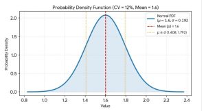
Which I find a bit sobering: it’s not very accurate even if you believe it’s worth measuring in the first place!


